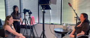
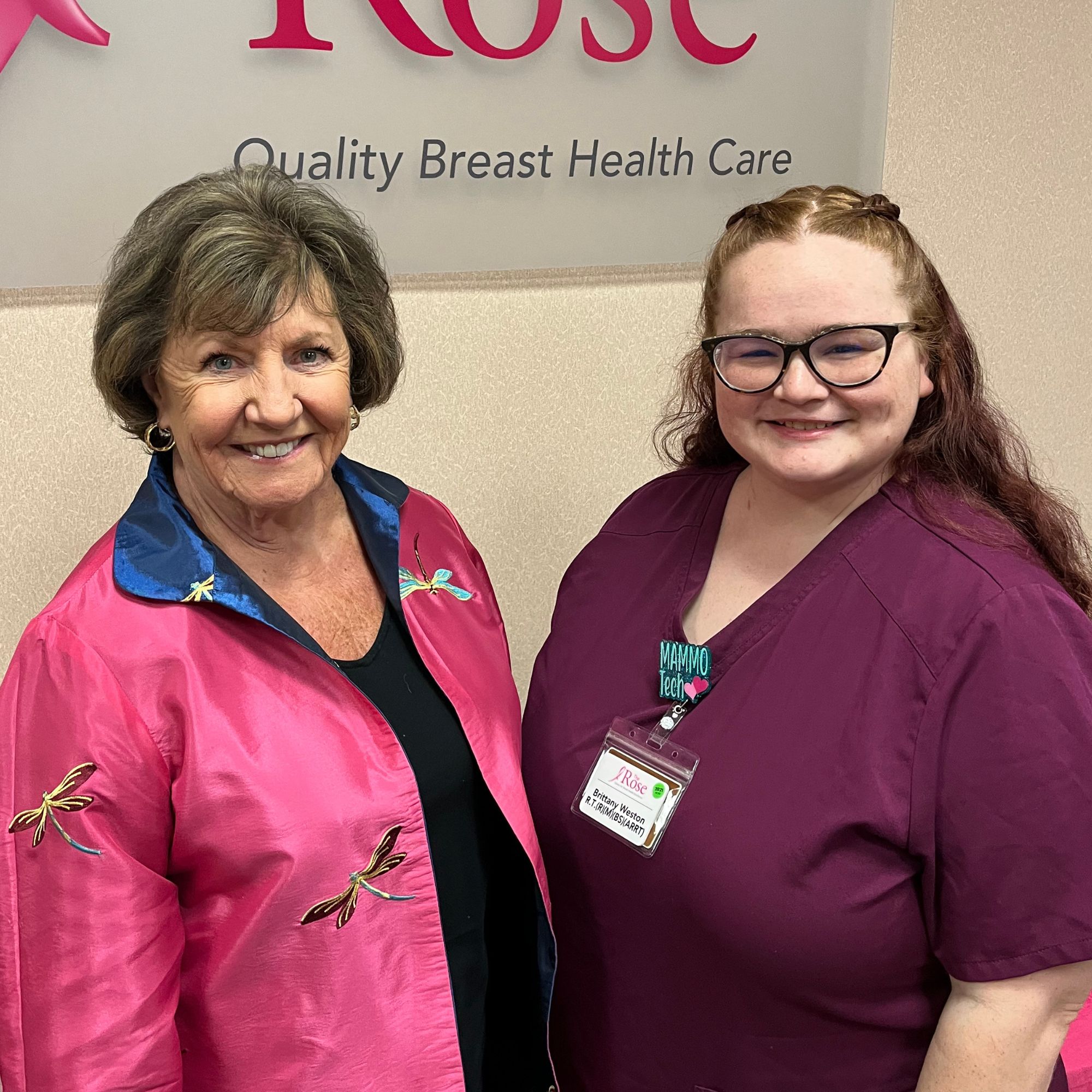
Dorothy: [00:00:00] Right after we launched the podcast, one employee after another told me that I needed to interview Brittany Weston. Finally, we talked her into doing this. And I have to tell you, Brittany is not only an excellent mammographer, but she’s also one of the best ultrasound techs we’ve ever had, and she assists in stereotactic biopsies.
During this episode, you’re going to learn how she helps women calm down during their imaging and how she talks with them to get them through a really scary time. Brittany began as a student at the Rose and is now one of our lead mammographers. During her time in radiology school, her mother and her grandmother were diagnosed with breast cancer.
So this job isn’t just a career, it’s a purpose.
Let’s Talk About Your Breasts. A different kind of podcast presented to you by The Rose, the Breast Center of Excellence and a Texas treasure. You’re going to hear frank [00:01:00] discussions about tough topics. And you’re going to learn why knowing about your breast could save your life. Join us as we hear another story and we answer those tough questions that you may have.
Brittany: My name is Brittany. I am one of the lead mammographers here at the Rose. I also do ultrasound and bone density as well.
Dorothy: So Brittany, tell me why everyone said when we started this podcast you have to interview Brittany.
Brittany: The Rose is very important to me. When I started here as a student, I knew that I wanted to work here.
I knew that everything that it stood for, I knew Dr. Melillo, from Dr. Raz, to all of our other doctors were some of the greatest doctors that I’ve ever met in my time. And I met a lot of doctors. So, I just knew I want to work here with all the girls that were working here at that time and was training me.
Dorothy: You came to the Rose as a student. Now, what does that mean exactly? Were you, you were not… [00:02:00] Were you in radiology school or before that?
Brittany: I had finished radiology and, um, I decided to further and go into multimodality, which I chose mammography because it was something close to me. My grandmother had been diagnosed while I was in x ray school.
Oh, and so, and then during mammography school, my mom got diagnosed. Oh my goodness. And so, I felt like that was it for me. That was why I was choosing this path. It was something that was going to be close to me in the future, and so God led me to choose mammography.
Dorothy: My goodness. Now, as a radiology tech, though, you could have gone to work right away in a hospital, correct?
Brittany: I did, kind of. I was doing, uh, part time at Memorial Hermann, but it just, I… thought I was going to love it a lot more than I did. And so when I started mammography, I felt an instant click with just what I was doing and, and what I was learning that I [00:03:00] enjoyed it so much. I knew x ray wasn’t going to be for me, but I think mammography was going to be. What I chose.
Dorothy: So when we’re talking x ray, we’re talking chest x rays or, uh, broken bone x rays, or you, you could have done any of those things, but you decided to specialize in mammography. And what does that mean exactly?
Brittany: So it means that you’ve done your special, you’ve done your x ray, uh, and then you’ve chosen to do something that will add to your, add to your experience and your education to further your career.
Dorothy: So you’re a licensed, uh, radiology technologist first. What does it take to become a mammographer?
Brittany: The way I did it is I ended up going through schooling. So I did my x ray and then I chose to go into another semester of schooling.
So I got to, uh, stay in that program kind of, but go to mammography and learn about how to do it. And we did clinicals and we did [00:04:00] in classroom and we learned all about the stuff that we wanted to, uh, with everything to do with mammography.
Dorothy: And so when we talk in clinical, does that mean you observed or you actually did it or what is that?
Brittany: Yes, so clinicals were, we came to wherever we were assigned and I was assigned here at the Rose as a student. So I came in and I I saw a couple and then I was told, okay, go for it. And so it’s kind of like being thrown in the deep end, but it was nice because it was like, you actually got to have more hands on.
And so I liked doing the clinicals.
Dorothy: Right. And you’re supervised the entire time, of course.
Brittany: Yes.
Dorothy: So, yeah, that had to be nerve wracking too.
Brittany: It was, especially because. This, this, this modality is so important that you get your best imaging. And as a student, you’re not, you’re your best yet. Um, so it’s like, okay, what else do I need to do to get this better image, to get this great [00:05:00] image that I can get to these doctors to have this patient get the best quality.
And get the best outcome and I didn’t want to ever mess that up. So I was always striving perfection.
Dorothy: Right now and this is something I don’t think we’ve ever talked about in any of our uh discussions Every woman’s breast is different.
Brittany: Oh, yes.
Dorothy: And every woman’s positioning is different, but you have to have certain angles that that are absolute.
It’s like you have to have them. So can is it possible to describe that at all?
Brittany: So we always want to see different angles because there’s different things that can be given in those different angles. So when we do what’s called our CC view or our top down image, we’re looking going anterior to inferior or top to bottom.
Um, and so we get a good overview of what that breast is going to be. And it gives us, cause we go off of like a clock face. So if we think about a clock, if you fold it in half, That’s the way [00:06:00] your breast is on that view. So when we go to our other angle, which is our MLO angle, we get to see the clock as kind of like a turn to the side and then it all matches up and it makes.
This perfect image basically of where things are located. So you can have something on one side of the breast in one image, and then when you turn it, it goes to another, and it all kind of works together to find what’s hiding in your breasts.
Dorothy: So, do breasts, are they kind of similar? I mean, you might find something in one, but you wouldn’t see it in the other.
Is that a flag?
Brittany: Um, it can be. Well, what I would say we would look for is, Some things are going to be normal, so there are certain things like vascular calcifications, which is like your vessels and your arteries you’re starting to calcify that that can be more in one versus another, but there are red flags that we see those calcifications that look a little [00:07:00] bit different or a mass.
that looks nice and round, or if we see something what’s called speculation, it looks like a star. It’s, those are the red flags we look for, and as a tech, if we see those, we like to go present them to the doctor as soon as we can.
Dorothy: And can you see something that a woman doesn’t even know it’s there?
Brittany: Absolutely. Half of the time our patients don’t feel anything. Um, they could say they have lumpy breasts all the time, but Unless they’re actually doing like a good breast exam, something that feels different isn’t going to be, no.
Dorothy: Mm hmm. And sometimes it can be so small or like calcifications. That’s not something you necessarily feel at all, right?
Brittany: That’s why screening mammograms are so important.
Dorothy: Yeah. Yeah. It spots it before we can ever feel it.
Brittany: Yes.
Dorothy: And so do you have women that are, are real difficult to image?
Brittany: It, I think it all [00:08:00] depends on who you’re, who’s doing the imaging. If they’re comfortable with you, they’re more relaxed. The more relaxed they are, the better the images are going to turn out because they’re putting their trust in you.
And it is, it’s a very vulnerable position for a woman to be in. And it’s very important that they make, that you make them feel as comfortable as they can be. And so…
Dorothy: Now how do you do that? I mean, here I am a woman and I am… Without any clothes on, and you are moving me around this machine, how do you make a woman feel comfortable?
Brittany: I like to ask them, are you okay? Are you doing okay? I like to let them know that no matter whatever happens, we’re going to be here for you 100 percent of the way. Especially when they’re coming in for those diagnostic exams, they come in and they’re nervous and they’re, they’re scared out of their mind.
What’s going on? What’s, what am I feeling? Why am I being called back? And you just let them know, even if insured, uninsured, it doesn’t matter. You let them know, we here at the Rose, we’re going to be here for [00:09:00] you. We’re going to be every step of the way. Start to finish, if you ever need anything, give us a call, we can help.
Dorothy: We all know this happens. I have been on that table. You’re looking for something, and I do want you to talk about ultrasound and the difference between it and mammography. But you’re looking for something, and you know you saw something. How do you keep the patient calm? Or how do you… not express that concern on your face.
Is that a, is that something you learn? Is that something that’s natural for you? How does the tech do that?
Brittany: It’s definitely harder some days than others, especially when they’re telling you your their story. It’s definitely control. You have to learn. It’s never something that’s a good feeling like, “Hey, I found this thing!”
It’s “Oh, I found this thing.” You try to let them know that no matter what it is, anything has to be tested before it can be for sure.
Dorothy: We can never tell cancer just from a film.
Brittany: You can tell that it looks different and you [00:10:00] can, you can have more suspicion than others. But until we get that pathology back that says for sure what it is, there are a lot of different things that can mimic a cancer and until we get those specimen back.
So usually I just, I let them know, the doctor’s going to explain everything to you. They have, they have more imaging, they have more experience. So the doctor is going to be the one to ask all your questions. I can answer as best I can, but any real questions that you have or any concerns that you have, our doctors are some of the best that’ll answer those questions.
Dorothy: And as a technologist, are you used usually in the room when the woman is talking to the doctor?
Brittany: Absolutely. Every time we have a doctor come in, we always have a technologist in the room just so because sometimes our doctors use big words or they try to explain it. I like to go back and I like to say, okay, so this is really like a little bit less what it means.
So they say this speculated mass. Well, it looks a little like a star or [00:11:00] it’s calcifications. Well, it’s calcium deposits. It’s like little grains of salt or it’s like you have dense breasts. You’ve, you’ve like live, look into the clouds. You see a cloud, you have breasts that can hide things. It’s not necessarily bad.
It’s just different. And unfortunately, Sometimes there’s still those nerve wracking and like, well, what does that mean? Is this what this means? What can you tell me? I’m like, I can’t tell you anything, but I can explain everything that is going to happen.
Dorothy: Yeah, well, that’s a good way to put it. So now tell us why would we have to have an ultrasound after we’ve already gone through this whole mammogram thing?
Why do we also need an ultrasound sometimes?
Brittany: So ultrasound and mammography are like sisters, they work really well together. They, you have these denser breast tissues that can hide things, you do your best to get everything you need on mammography. But sometimes that ultrasound is just going to help you out a little bit.
It can hide smaller things, it can hide tiny [00:12:00] cancers that can’t be seen on mammogram yet. Or it can hide cysts that can get large and painful and it can hide tiny cysts. Um, it allows us to see. In the ducks that we can’t see in a mammogram. It allows us to see, um, lymph nodes. It allows us to see whether or not you’re having reactions, infections, all of that.
It works together with the mammogram so well. It’s why a lot of our dense breast patients, they come in for both because they’ll work so well together to create an entire story of the breasts.
Dorothy: So you can really tell infections and things like that on an ultrasound?
Brittany: Oh yeah. Uh, so we have a lot of patients that come in and they have redness or swelling, which can also be a certain type of cancer.
But if we come in and we see that there’s a lot of blood flow to that area or if there’s a certain amount of one area versus another, we can see fluid collection [00:13:00] with infection. And so we look at that and we say, okay, we can see if we can test some of this and see what type of infection it is, what antibiotics will work best for it.
And so we work, it works really well with that ultrasound to do both.
Dorothy: So what is the, how was the youngest patient you ever did?
Brittany: Um, so as an ultrasound tech, my youngest patient was 13. She came in and she was feeling a lump. and luckily it was nothing. It was just fatty tissue. But Even as a 13 year old, they’re doing breast exams, which is so important because I believe our youngest patient here was 21 who was diagnosed.
Dorothy: 19, actually, yeah. Before your time a little bit, but yes.
Brittany: I know, but I’ve heard like every, I’ve heard about all these patients that come in, they do, they get it so young. Yeah. And so, um. 13 was my youngest for ultrasound.
Dorothy: How did you calm her down?
Brittany: Um, luckily her mom was in there too. [00:14:00] So, that’s kind of like calming two people down.
Um, it’s like, because mom’s of course so nervous for her daughter and then daughter’s just nervous in another one. And so, it’s kind of just letting her know, okay, well, it’s a good thing that you’re doing your breast exam even though you are so young. It’s good that you know the difference in your breast and different feelings.
So, it’s praising her and letting her know she’s doing a good thing. It’s also letting her know or talking to her about school or talking to them about what they like to do on the weekends. It’s trying to take their mind off of it while you’re doing that exam.
Dorothy: You said that your mother and grandmother, grandmother were both diagnosed so.
How was their experience?
Brittany: So, my grandmother’s actually wasn’t found in her breast. Hers was found in her stomach. And so, they’re currently doing the anti estrogen and to help with that. Um, but hers is metastatic. So, they’re just trying [00:15:00] to treat everything that they find. For my mom, it was a little bit different.
Uh, she actually found hers when she felt her lymph node. And her lymph node was severely swollen and her doctor says, well, take these antibiotics for two weeks. It didn’t work, of course. Um, so she went back and they said, okay, you need to go get a mammogram. Well, they sent her with the wrong order. They sent her with a screening mammogram order.
And so then she had to come back again with a diagnostic order. And so when they came in for a diagnostic, they saw that she had a five centimeter lymph node. And, um, they said, okay, yeah, let’s do a biopsy. So, uh, they have found another area as well. So they did a biopsy of the lymph node and then the mass that they found in her breast.
The mass was like three millimeters. So super tiny. Uh, it turned out it was invasive ductal carcinoma. And she was grade three, I believe. Um, so she [00:16:00] went and she had chemo first. She had what’s called the red devil. Um, and so she went through that. It was a hard time for my mom. It was a hard time for all of us.
Um, but she got the chemo. After the chemo she had her, she opted for a double mastectomy because she didn’t want to go through it again. She wanted to make sure that it was gone. So, she had her double mastectomy and she had the latissimus flap.
Dorothy: Right, right.
Brittany: Um, and then, uh, after about, I think it was two weeks after her surgery, her side that she had her breast cancer on started rejecting the flap.
Dorothy: Oh, no.
Brittany: Yes, so it started, her, it started to reject and it started to die. So, she went back into the hospital and that can happen after radiation. So, she had had radiation. And it had rejected once she had that surgery because the radiation just kind of [00:17:00] killed everything there. So then she went back in, had it removed, let it heal, and then went back in for another surgery and had an implant placed.
And so her journey finally, she’s at her five years now.
Dorothy: Oh, super great.
Brittany: I had her switched from Memorial Herman over to the Rose.
Dorothy: Oh, great.
Brittany: Yes. Um, but her doctor had even told her, well, you don’t need to go get anything done anymore. I said, no, that’s, that’s not how that works. I said, we’re still going to get you going. We’re going to, unfortunately we’d have seen reoccurrences.
So we’re going to keep imaging and mammo ultrasound, everything.
Dorothy: Right. That had to be tough. You were pretty young.
Brittany: I was, I was, I think I was 20, 21, 22. My brother was 13, 14. And my sister’s was 25.
Dorothy: So did everybody look at you for all the answers?
Brittany: So [00:18:00] at that point, I didn’t know a lot. Yes, I was learning. I’m like, okay, what do we do now?
I was doing extra research and be like, okay, I’m gonna ask my teachers. I’m gonna ask people at the Rose. I’m gonna ask everybody. I know. What do I do? What do I do? Um, a lot of it was just being there for her. And just as she was going through it.
Dorothy: So do you ever see your mom when you’re doing someone else?
Do you ever think about her while you’re doing a patient?
Brittany: Oh, absolutely. It’s part of the reason that I let those patients know. I said, I’m going to give you my best exam. You don’t know why? Because if you were my mom, my sister, my grandma, I want you to have the best possible outcome. And to do that, I’m going to get you the best possible imaging.
I see them in every patient I do.
Dorothy: So you’re unusual because you can do both mammography and ultrasound. It had to be, is it certified for ultrasound or how does that work?
Brittany: So, um, I ended up doing cross training here at the Rose because during COVID we ended up not having as many techs. I’m like, well, let me learn something new I can help out.
So I decided to [00:19:00] learn breast ultrasound. And so. I went through the cross training certification, which is doing 200 exams, doing our continuing education credits, and then I went ahead and I took the test through ARRT. I got to pass, and so I am officially certified to do breast ultrasound. furthered my education not in a school setting but in a clinic setting.
Dorothy: But now that, that is not an easy test. I mean it includes so many different parts and it’s pretty detailed. Yes. I hear everyone talking about oh yeah I’ve just passed and I’m thinking my gosh that’s not a, that’s a huge test to pass.
Brittany: It is. It’s learning the physics of ultrasound. It’s learning your anatomy, your physiology, your what are you looking for, what do you measure.
It’s, it’s a very hard test. Ultrasound is one of the hardest modalities because it is very tech dependent. And if a tech misses it, then it’s really important.
Dorothy: You know, it’s, I know it’s hard to [00:20:00] explain when you’re just talking about it verbally, but what does that mean? It’s tech dependent.
Brittany: So Tech dependent is basically that the tech is doing all of your scanning.
Some of our doctors will back scan and they’ll look and they’ll feel and they’ll make sure that everything’s correct. But with ultrasound, it’s very tech, the tech dependent is the tech is going to find something. The tech is going to be the one who notices it because you don’t have the doctor in the room at that time.
Dorothy: And sometimes just the way you hold your wand can make a difference. Is that true or is that one of the myths?
Brittany: That is true. No, that is very true. We do different angles and we see it in different angles. So if you’re holding your probe backwards, it’s going to look like it’s on the opposite side when really it’s on the left side versus the right side.
Or if you have it at an angle, it’s actually a different view. And so it’s not the correct angle you want it at. So you want to make sure you have your probe straight whenever the view you’re doing.
Dorothy: [00:21:00] So ultrasound is actually explain that to me, sending a wave.
Brittany: Sound wave.
Dorothy: Sound wave through tissue.
Brittany: Yes. And it’s reverberating off of it and creating your image.
Dorothy: Okay. And that can show you if you have a cyst or a solid area or…
Brittany: Absolutely.
Dorothy: And I know, uh, personal experience here when you were doing mine and, and the doctor was in the room and she said, Oh, you’ve got the calcifications there. That’s not easy to find on ultrasound.
Brittany: Calcifications are not easy to find on ultrasound.
Dorothy: So how’d you do that?
Brittany: Um, it takes a trained eye. It is, it’s very, You have to be very focused on finding those because it’s kind of just like a blip of a bright spot. And so when you have a grouping, then it’s a little bit easier to find, but it’s still, you have to find it within all of that tissue and it’s gray and white and black and it’s all working together to create your image.
So finding those can be. A little bit of a challenge.
Dorothy: And you’re, you’re [00:22:00] correlating, how do you say that, with a mammo you’re trying to?
Brittany: Yeah, we’re, we’re using it in correlation with it.
Dorothy: Yes, and so you, you, but those are different, totally different dimensional things.
Brittany: Yes, but working together they create that perfect.
Dorothy: Still, you have to see that in your head.
Brittany: Absolutely.
Dorothy: Okay.
Brittany: It’s nice having the mammogram up and having it visualized while you can do your ultrasound. And just like with the mammogram, we use clocks with ultrasound.
Dorothy: Wait, how do you do that?
Brittany: We use the mammogram. We have the clocks. We say it’s at three o’clock.
So if you think about it, when you’re laying flat, your breast is more of a circle. So you start at 12 up and then you go around the clock and you have your one o’clock, your two o’clock.
Dorothy: Got it.
Brittany: And you find, so if the doctor says, I see these calcifications at 10 o’clock, I’m going to really pay attention to the 10 o’clock on that ultrasound as well to see if I can see them.
Dorothy: And, and is it [00:23:00] the same on each breast or…
Brittany: So, it’s, it’s the same on each breast, but it goes out. So on one side, it’s going to be three o’clock on the left. And then the other is going to be out on the right.
Dorothy: Ah, that makes sense. I mean, I think a lot of women, when they hear three and Six o’clock and all that, they were wondering what the heck are they talking about.
So, when you’re talking to women, and you see how many a day?
Brittany: Uh, it can vary. We see, each check can see between 12 to 20.
Dorothy: And do you find yourself giving one major piece of advice to each of the women?
Brittany: Come back. It’s very important that you come back.
Dorothy: It’s not a one and done.
Brittany: It’s never a one and done.
Dorothy: And why is that?
Brittany: Breasts are forever changing. You gain weight, you can lose weight, it’ll change your breast tissue. You can take estrogen and progesterone and as a younger [00:24:00] patient that’s going to change with birth control. And then hormones can change your breasts, periods, and everything like that. It’s, you’re forever changing.
And so it’s always important that each year you do your exams. But if, also you’re doing your breast exams at least once a month. Because if you feel something new or different we want you to come in. We don’t ever want you to wait until it’s your yearly. Because you can change in two months.
Dorothy: Oh, yes. Yes, I think that’s a big message.
Right? Women go, I just had my mammogram. It can’t be anything.
Brittany: Oh, yeah. Yeah. Unfortunately, we’ve seen that as not true.
Dorothy: Right.
Brittany: So it’s always important to do those breast exams.
Dorothy: And the other thing I think we particularly are concerned about is young women who have just had children. We see a lot of cancer in that age group, and is it harder to image when, when you’ve just had a child?
Brittany: So, it depends. We do, for patients who are lactating, we do recommend that they pump at least [00:25:00] 30 minutes prior to their exam because we wanted to get the best possible image. And so, if they pumped, then we don’t see as much white or milk in that breast, so we get to visualize it a whole lot better.
Dorothy: Oh, that’s a good point.
Yeah, yeah. I wonder if doctors ever tell their… There are patients that before they come in?
Brittany: So we tend to do it. And our schedulers are aware, luckily that.
Dorothy: Yeah, they know how to.
Brittany: Yeah. They know how to give ’em good instruction.
Dorothy: What is your hope for women in the future? Brittany, you’re young, you, you’ve seen a lot.
You’ve been through a lot.
Brittany: I hope that they continue to do everything that they need to do, that they’re conscious of how they’re working. Because you know, women, we are strong. Mm-Hmm. . And we are very stubborn at times. To the point where we’re going to take care of everybody else first and then ourselves.
So I’m hoping in the future we learn to kind of delegate that and put ourselves as a priority with [00:26:00] everything. Cause it’s, unfortunately a lot of what we see is women who’ve waited because they’re taking care of somebody else. Or they’ve had stuff going on. Or they haven’t been back because their kids have been sick or they’ve lost a loved one.
And so it’s hard for them to put themselves first. So my hope is in the future that women learn to delegate and we can put ourselves as a priority.
Dorothy: So you’ve been with the Rose for five years and you came to us right out of school. You sound like you’re 50 years old. How old are you? Really?
Brittany: I turned 29 this year.
Dorothy: Ah, ah, such, so much wisdom to have for such a young person. Truly, Brittany, we’ve all admired you and appreciated you and really, you know, the way that you’ve always stepped up to the plate and said, I can do more, I can do something else. That’s so important. You are setting up a career that could last you a very, [00:27:00] very long time as, as the modalities change.
I mean, are you ever, or have you even started thinking about stereotactic biopsy? Are you doing that yet?
Brittany: So, uh, I actually started doing stereo my first year.
Dorothy: Wow. Wow. I didn’t, I don’t think I’d put that together.
Brittany: Yeah. So I, I do know how to do the stereotactic biopsies.
Dorothy: And what do you love the most?
Brittany: Um, I love mammogram.
I do love doing mammograms. I love my ultrasound and I love doing bone densities and all the other stuff that I am capable of doing, but I love I love being that imager for a patient who’s coming in that’s worried or a patient who’s coming in for their annual or I just, I love being able to get those images that’s going to give them the best possible outcome.
Dorothy: And so if you had one word that would sum up everything the Rose is and all it means to you, what would it be?
Brittany: Family.
Dorothy: Ah.
Brittany: We’re family here.
Dorothy: Yeah.
Brittany: I mean, we start from one end work to the other where [00:28:00] we’re just, we all work together and I just love it about us. I love that. We’ve worked here or they’ve worked here for so long in that no matter what, what a new person comes on.
They’re automatically family.
Dorothy: Oh, that’s good. That’s good to know, yeah. I know, you know, I, I, I hear, uh, sometimes I say, well, we can disagree just like families, but we can certainly take care of each other just like families, and I, I think that is important. Thank you so much for being with us. Any last words to young women getting into medicine?
Brittany: Persevere. You can do it.
Dorothy: Good, good advice. Well, thank you for being with us and for sharing so much. I appreciate everything you do and I can see why everyone says you need to interview Brittany. Thank you for being with us today.
Brittany: Thank you.
Dorothy: So that wraps it up for today and don’t forget we’re doing a episode every single day.
You’re going to get your [00:29:00] daily dose of Let’s Talk About Your Breasts during the month of October.
Post-Credits: October is the month of pink. And for The Rose, a Breast Center of Excellence, that means we’ll be airing podcasts every day in October to celebrate Breast Cancer Awareness Month. You’ll be hearing from longtime firefighter, senior captain, and breast cancer survivor from the Houston Fire Department.
You’ll hear how making clay heads was part of her treatment. Be sure to share with family and friends, because there’s a little something for everyone. To find out ways to help The Rose, visit our website at therose.org. And remember, self care is not selfish. It’s essential.
